Cardiac Rhythm Device Troubleshooting
1
2
3
4
5
6
7
8
9
10
11
12
13
14

  
HeartRhythmBox, an education page of heart rhythm topics.
visit us at www.heartrhythmbox.com
or https://www.facebook.com/HeartRhythmBox/

  • SETTING
    Pacemaker
  • GENERAL DIAGNOSIS
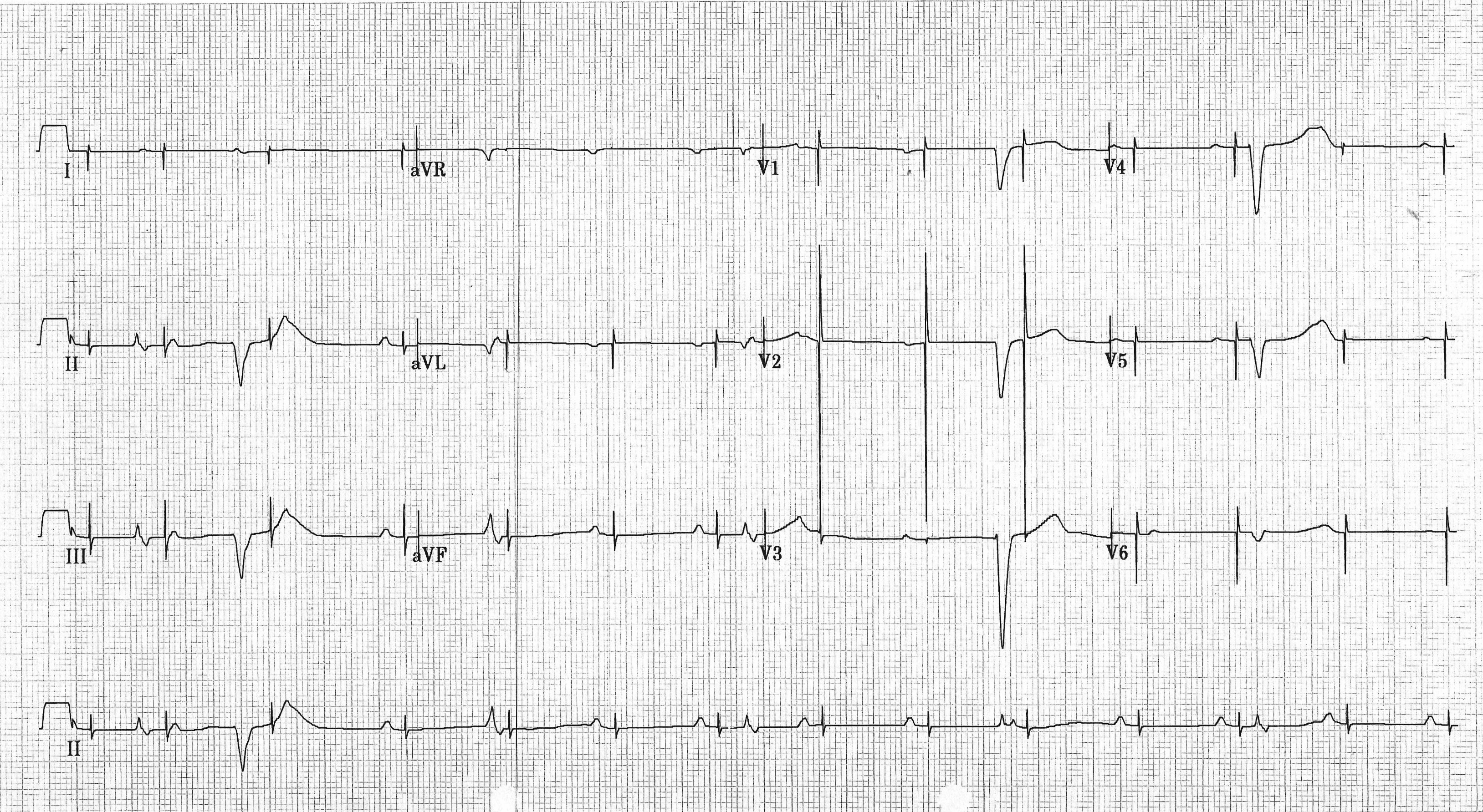
    Undersensing both A & V
  • EXPLANATION
    - Dual chamber pacemaker (2 pacing spikes within 240ms of each other).
    - Baseline rhythm: complete heart block with slow and narrow ventricular escape.
    - No evidence of appropriate sensing either A or V. Pacing spikes are all over the place.
    - Hard to evaluate pacing function. All pacing spikes occurred during functional refractory periods.

  • SETTING
    DDD for complete heart block x 10years; generator change 3 years ago.
    Presents with syncope while pulling a field tent. Initial pacemaker interrogation shows normal pacing parameters.
  • GENERAL DIAGNOSIS
    Failure to Fire/Pace
  • DIFFERENTIAL DIAGNOSES
    • Oversensing: cross-talk, EMI, oversensing of P or T wave.
    • Lead problems: fracture, insulation break.
    • Circuit failure
    • Battery failure
    • Loose set screw
    • Incompatible lead & header
  • EXPLANATION
    - Syncope occurred during shoulder activity. Pacemaker should be checked during arm/shoulder movement. In this case, lead noise is observed during arm movement. Lead fracture is the root cause of the problem.
    - Loose set screw and header/lead mismatch are more likely to occur early after implantation or generator change. - Pacemaker was only 3 years old. Battery depletion is unlikely.

  • SETTING
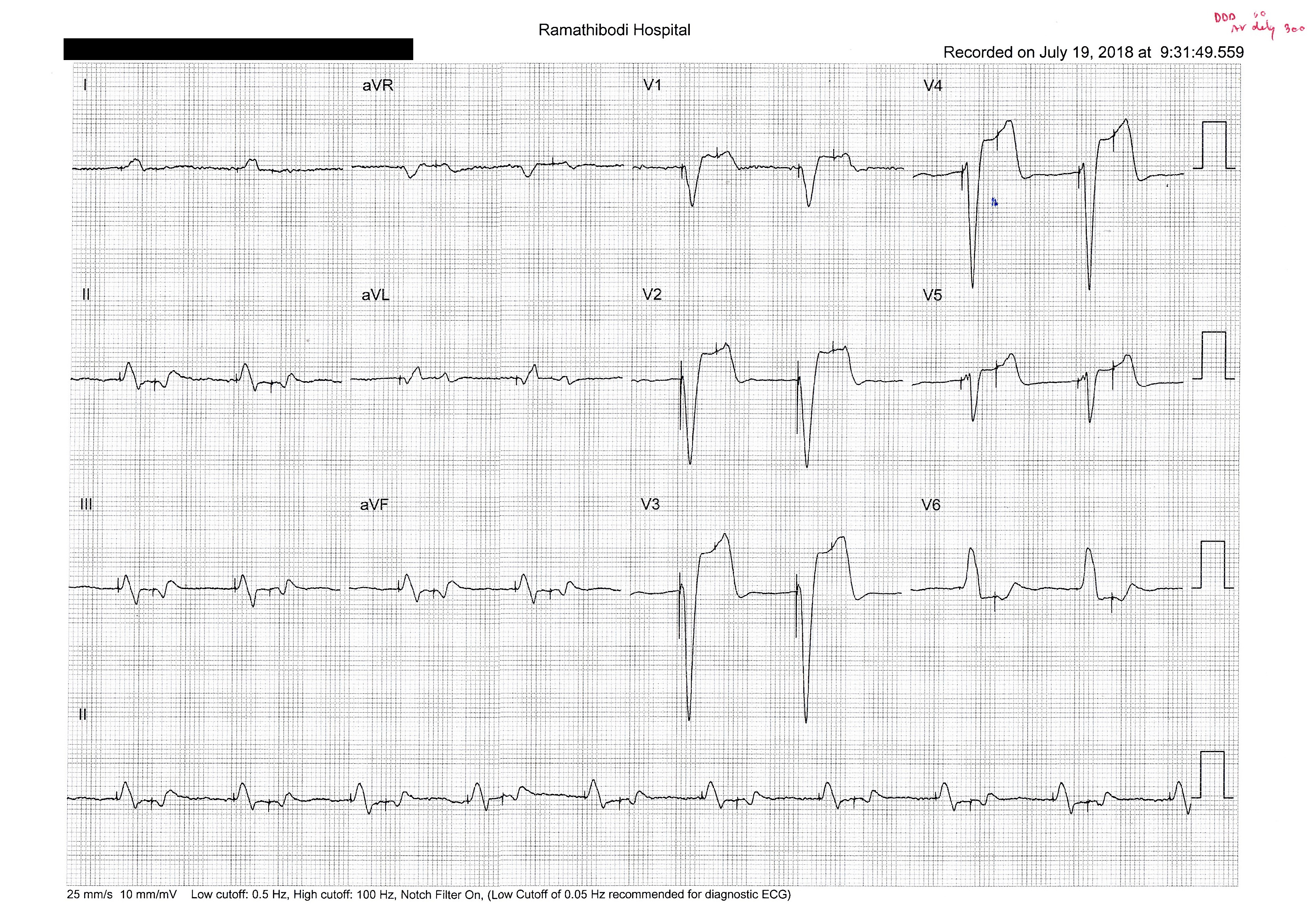
    DDD 60/120, AV delay 200. Tracing obtained 6 hrs after the implantation.
  • GENERAL DIAGNOSIS
    Failure to sense and capture of atrial lead.
  • EXPLANATION
    - DDx: Battery depletion, lead fracture, lead dislodgement.
    - The event occurred early after the implantation. Lead dislodgement is most likely.
    - In this case, the atrial lead was dislodged and not properly sensed or paced.

  • SETTING
    CRT-D; DDD 60/120; SAV delay 120ms; PAV delay 150ms
  • GENERAL DIAGNOSIS
    Loss of P-synchronous Pacing
  • DIFFERENTIAL DIAGNOSES
    • Long programmed AV delay
    • Atrial rate is too fast or maximal tracking rate is too low.
    • PVCs and/or PVARP extension post PVCs.
    • Atrial undersensing
  • EXPLANATION
    - Baseline rhythm is sinus rhythm, 1AVB, IVCD.
    - Only one pacing spike is observed at the 8th beat. The pacing spike is in sync with P wave and is biventricular (positive in V1 & V2).
    - Native PR is longer than programmed AV delay, so there should be biV pacing followed each and every P wave.
    - Intermittent atrial undersensing is the most likely the root cause of suboptimal BiV pacing.
    - In this case, A signal was tiny, recorded at 0.3mV.

  • SETTING
    Two years after dual chamber pacemaker implantation with dyspnea on exertion and lower extremity edema.
  • GENERAL DIAGNOSIS
    Failure to capture of atrial lead
  • DIFFERENTIAL DIAGNOSES
    • Lead dislodgement
    • Elevated threshold
    • Lead fracture
    • Insulation break
    • Loose set screw
    • Battery depletion
    • Circuit Failure
  • EXPLANATION
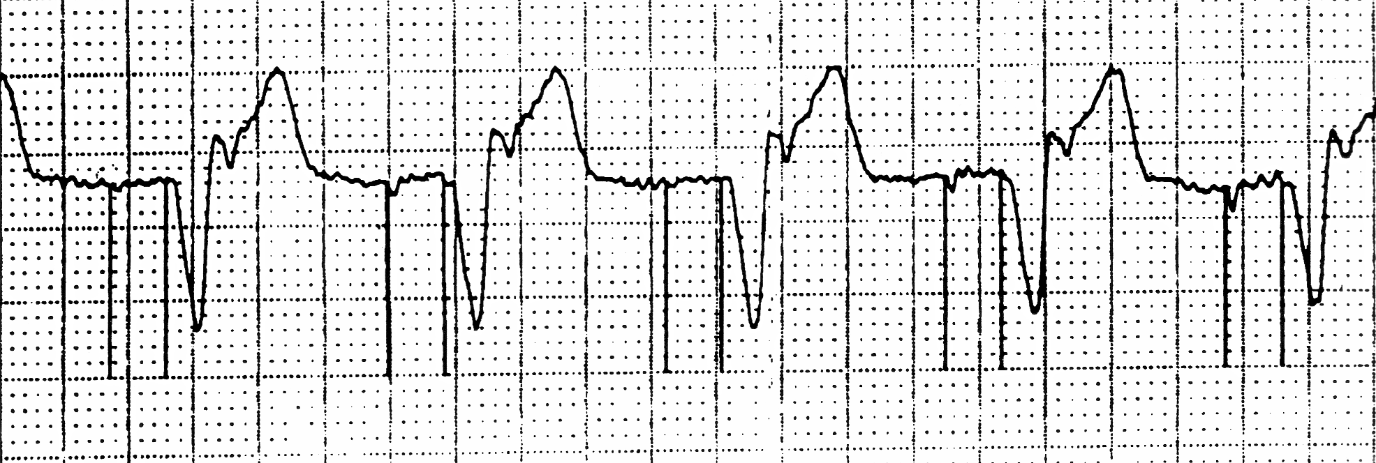
    • Tracing: 2 pacing spikes within 160 ms, no A capture, but V capture with retrograde P waves.
    • Retrograde P waves are within PVARP; did not cause PMT.
    • Symptoms of dyspnea and edema are likely from pacemaker syndrome from retrograde P waves.
    • Elevated threshold, rather than lead dislodgement, is more likely at the chronic stage of pacemaker.
    • Lead fracture, insulation break, or battery problems remain possible but less common.
    • In this case, the atrial threshold is up due to newly developed renal insufficiency.

  • SETTING
    DDD 60/120, AVD 200ms. One week after initial implantation.
  • GENERAL DIAGNOSIS
    Failure to sense and capture of ventricular lead
  • EXPLANATION
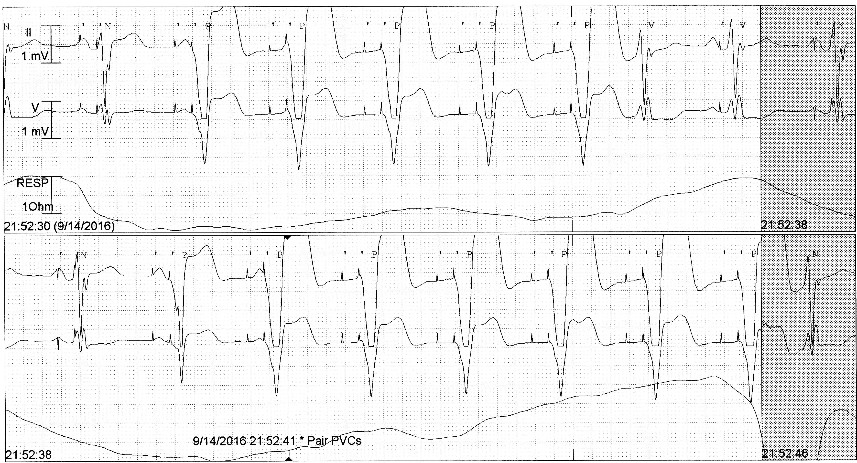
    • Atrial lead sensing function is normal, pacing function is unknown.
    • DDx: Lead dislodgement, battery depletion, lead fracture, insulation break.
      Since the event occurs in the acute phase, battery depletion and lead fracture are unlikely.
    • Baseline rhythm = sinus rhythm with complete heart block with slow ventricular escape.
    • Pacing spikes are P-wave synchronous. Some P waves are hidden in PVCs.
    • In this case, the ventricular lead is dislodged. Reposition is required. While awaiting repositioning, the patient should be managed as if he/she is having complete heart block without permanent pacemaker.

  • SETTING
    DDD 60, AVD 300ms, Boston Insignia. One day after initial implantation.
  • GENERAL DIAGNOSIS
    Failure to sense and capture of atrial lead
  • EXPLANATION
    • P waves (hidden in T waves) were not properly sensed.
    • A pacing resulted in V capture.
    • V pacing (the spike was 300ms after the A pacing spike) resulted in functional non-capture.
    • DDx: Atrial lead dislodgement in to right ventricle or leads switch in the header (A lead in the V port and V lead in the A port).
    • If A lead is in RV and A pacing results in V capture, should there be "safety-pacing" within 110ms?
    •    •In this case, it is the Boston Scientific device which has the algorithm to asynchronously pace at the programmed AV interval instead of safety pacing at 110 ms.
    • Can we rule in/out leads switch in the header?
    •    •With this tracing alone, cannot. To rule in, you need to see the evidence of atrial sensing. Though there is no such evidence in this tracing, it might be that we have not seen a long enough tracing.
    • Next step: PPM interrogation and CXR.
  • MORE INFO
    CXR and Device EGM shows atrial lead dislodgement into right ventricle.

  • SETTING
    DDD 60/120, AV delay 170 ms, tracing obtained in the recovery area right after pacemaker implantation.
  • GENERAL DIAGNOSIS
    Failure to capture of atrial lead
  • EXPLANATION
    • Magnet application (= DOO) shows that A pacing results in V capture.
    • DDx: A dislodgement in RV vs. leads switch in the header (A lead in V port and V lead in A port).
    • P wave from the native sinus beat is properly sensed meaning that there must be a lead in the atrial. So, this is the case of leads switch in the header. Reoperation is required.
    • Baseline rhythm = sinus with frequent PVCs.
    • After each sinus P wave, PVARP extension is triggered as a response to PVC.
    • Atrial pacing after sinus beat comes right on time of 830 msec (1000-170).

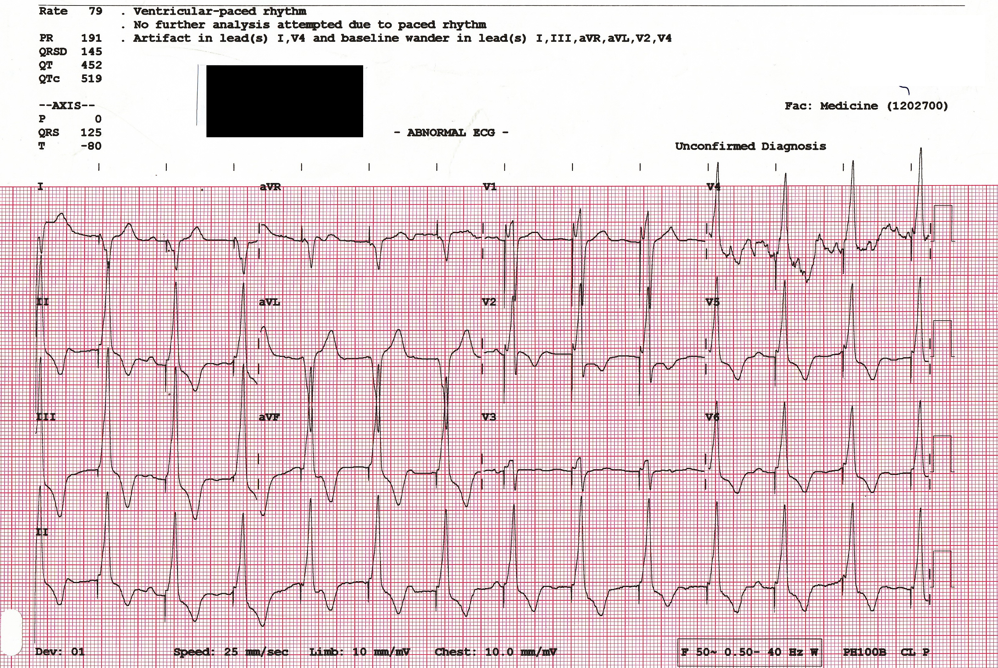
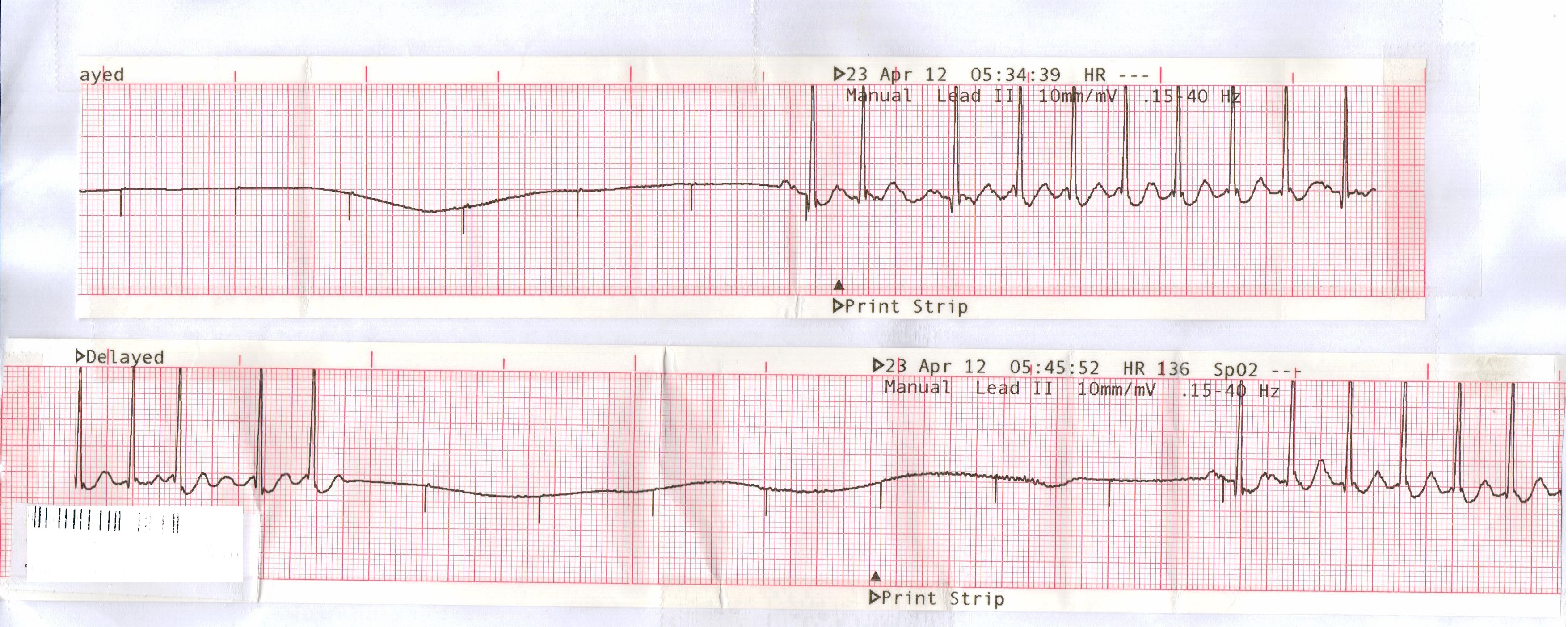
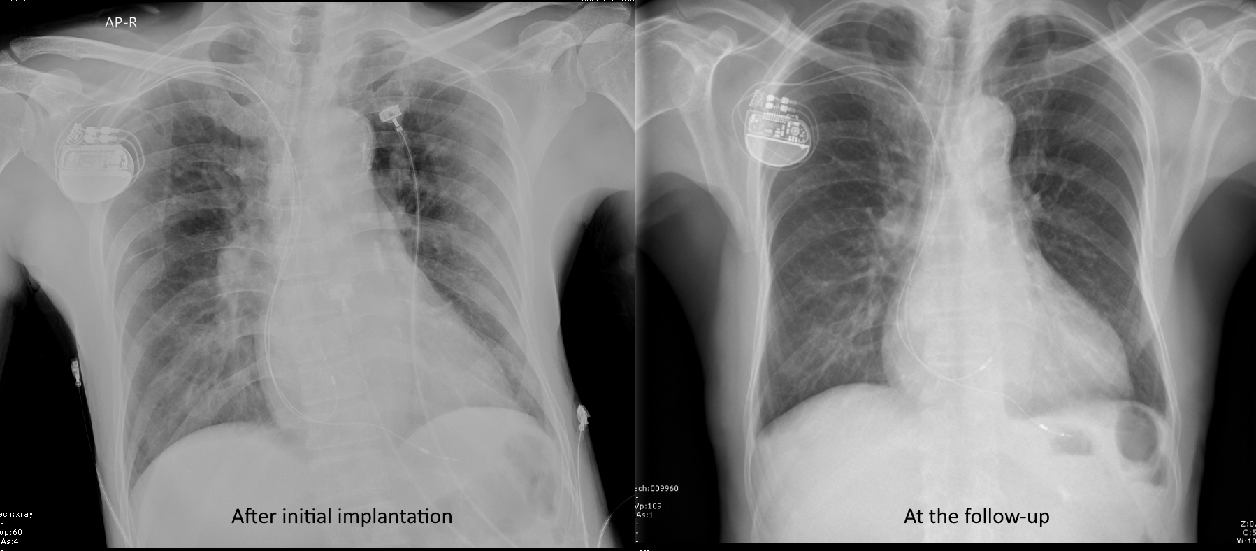
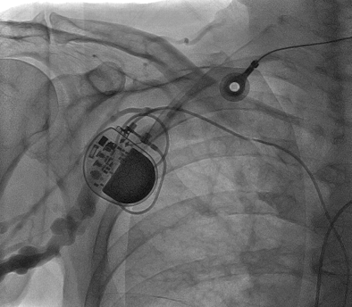
  • SETTING
    VVI 60. Initial implantation 15 years ago. Generator change 3 years ago.
  • GENERAL DIAGNOSIS
    Failure to output, failure to capture, and failure to sense.
  • EXPLANATION
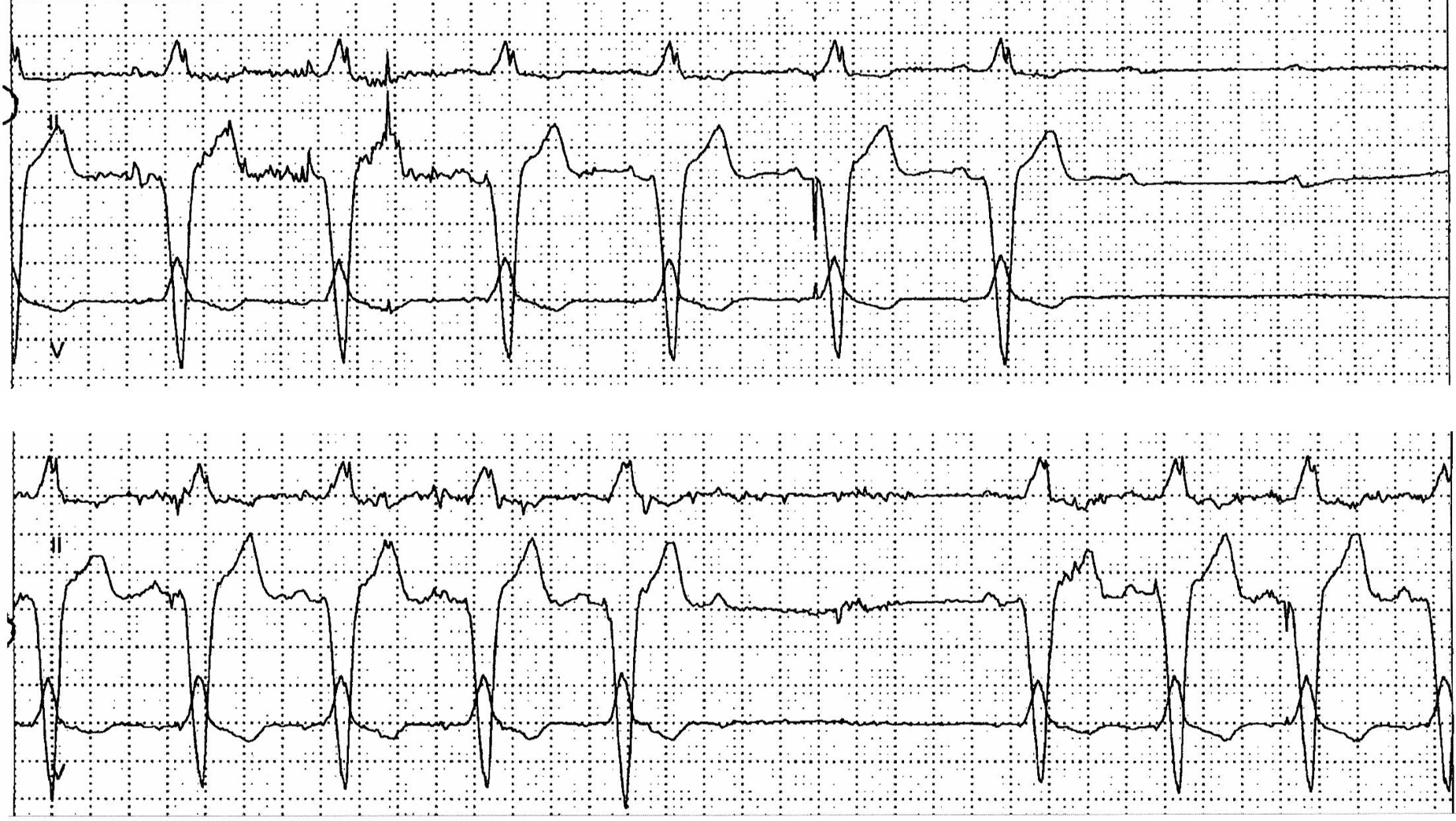
    - Baseline rhythm: AF with slow ventricular response; slower than lower rate of pacemaker.
    - No pacing spikes during the first four beats = failure to output.
    - 3 pacing spikes without capture at the last part of the tracings. The 1st and 3rd spikes are not in refractory period and are not captured.
    - Pacing spikes are not in sync with the native beats = failure to sense.
    - Lead problems are on the top of the list. Battery depletion is possible but less likely for a 3-yr old modern pacemaker.
    - In this case, lead impedance is 1800 ohms consistent with lead fracture. The fracture is also visible in the X-ray.

  • SETTING
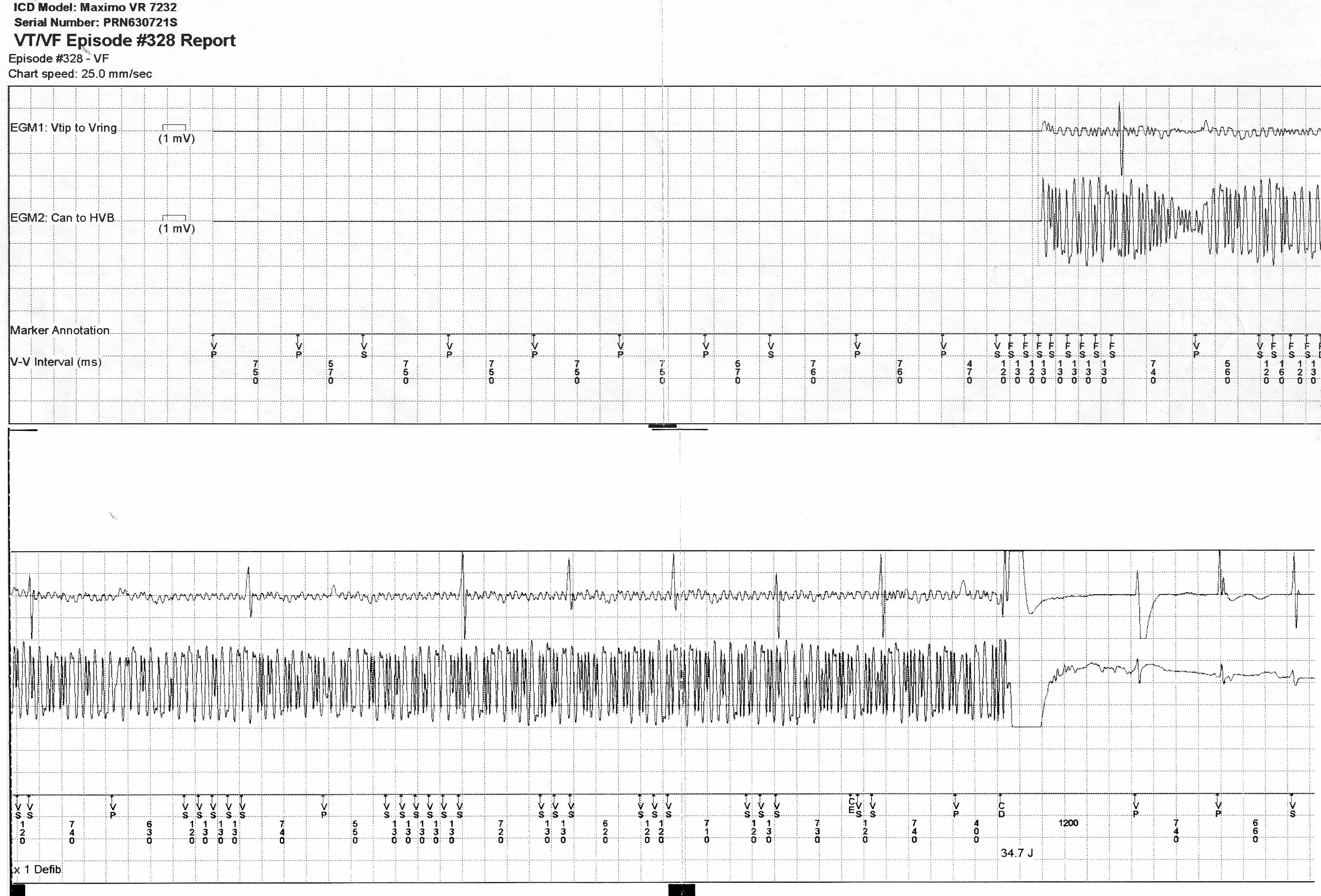
    Single-chamber ICD.
  • GENERAL DIAGNOSIS
    Inappropriate ICD shock
  • EXPLANATION
    - Signals with non-physiologic intervals (ie. <200ms) are recorded. These are noises.
    - Electromagnetic interference is most likely since noises are seen in both near-field (Vtip-Vring) and far-field signals (Can to HVB).
    - EMI causes inappropriate shock.
    - The patient reports use of electrical saw during the episode.

  • SETTING
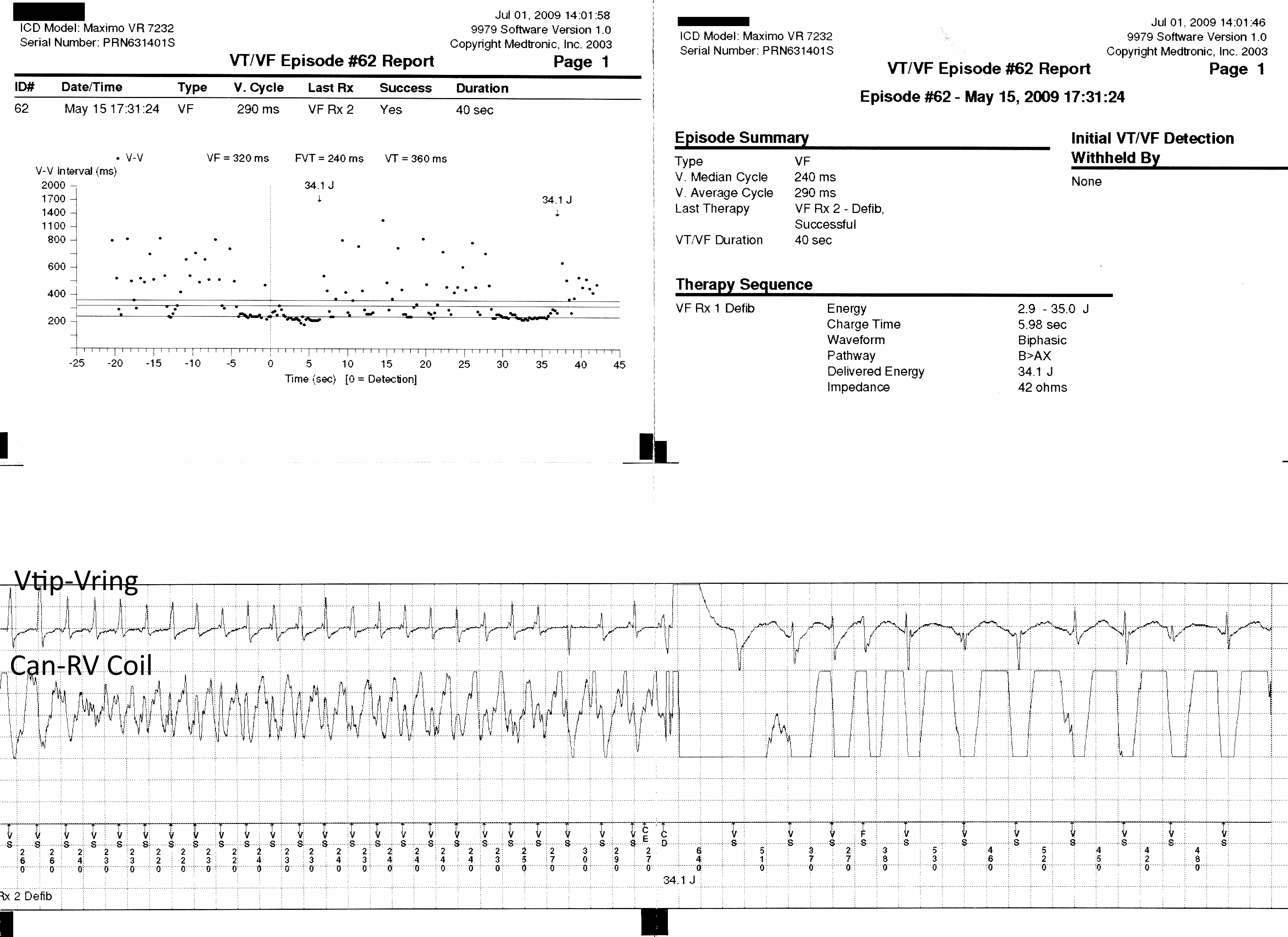
    Single-chamber ICD. Multiple shocks within 12 hours.
  • GENERAL DIAGNOSIS
    Appropriate ICD Shock
  • EXPLANATION
    - Both near-field and far-field signals are consistent with VF.
    - Multiple appropriate ICD shocks for ventricular arrhythmia within a short period of time should be treated as electrical storm.

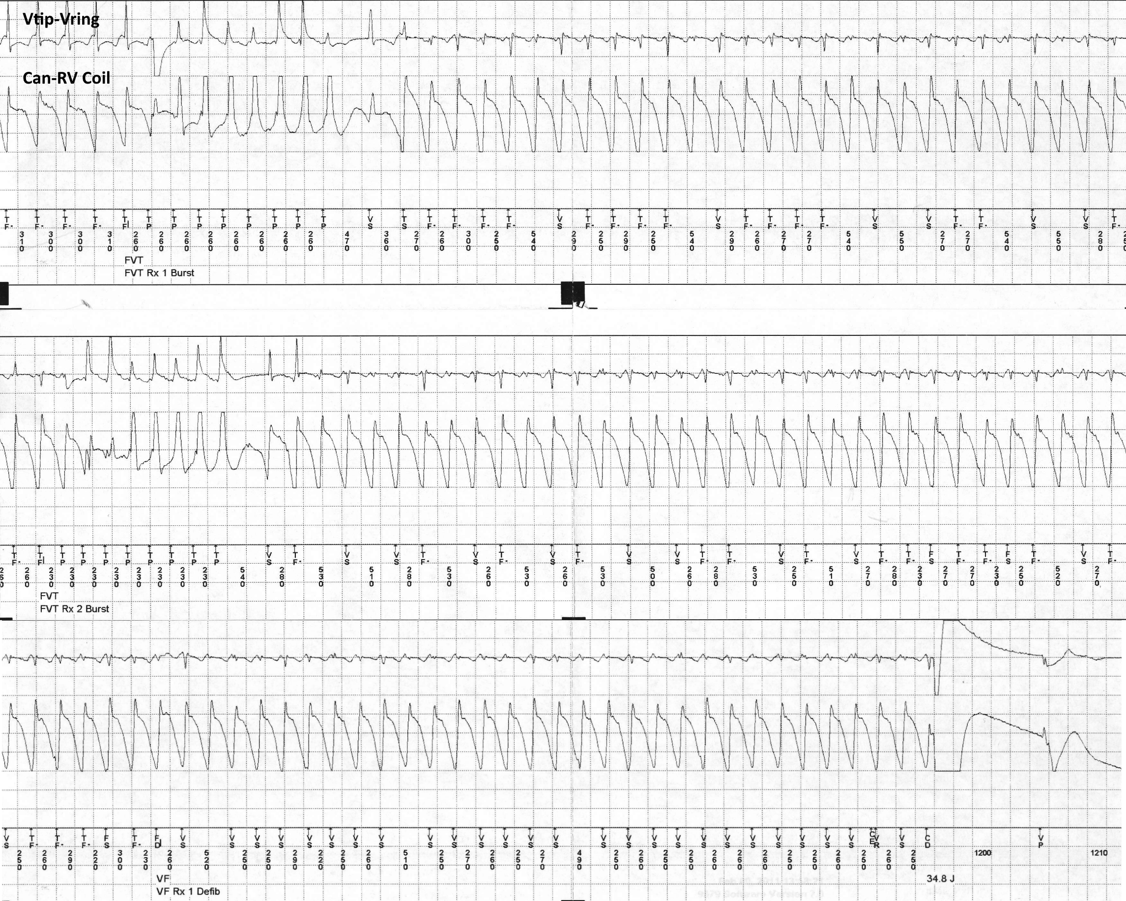
  • SETTING
    Single-chamber ICD. Palpitation and syncope.
  • GENERAL DIAGNOSIS
    Appropriate and Necessary ICD Shock
  • EXPLANATION
    1. VT with CL 300 ms triggers appropriate ATP (FVT Rx1 Burst).
    2. VT accelerates to 260 ms.
    3. Several VT beats are undersensed; noted with the CL of 500-540 ms.
    4. The second therapy (ATP) and the third therapy (shock) are delayed.
    5. Shock is necessary after unsuccessful ATPs.
    6. Ventricular lead sensitivity should be assessed.
    7. VT storm should also be treated accordingly.

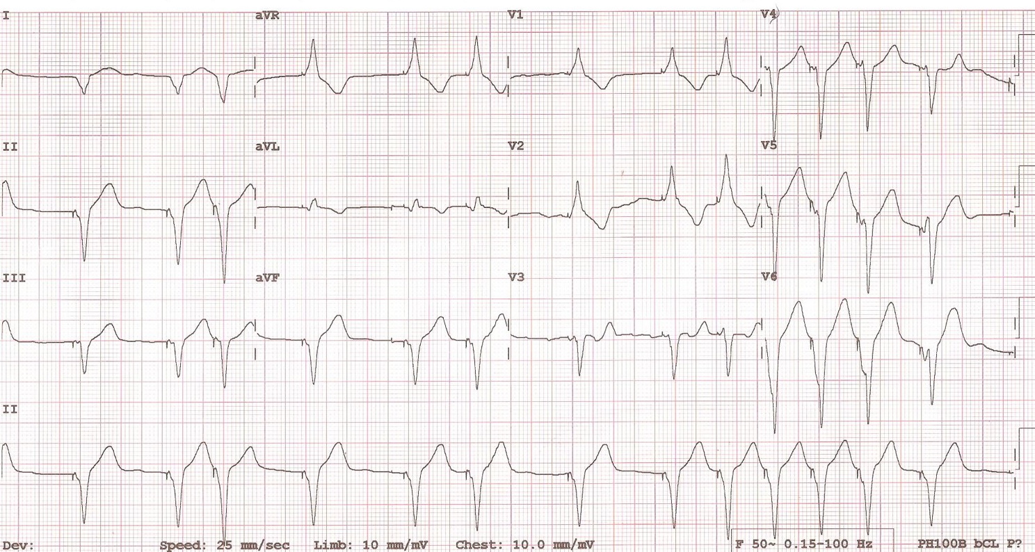
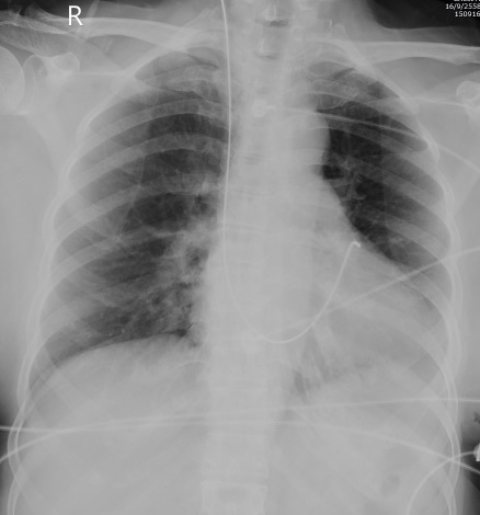
  • SETTING
    Temporary pacemaker. VVI 80.
  • GENERAL DIAGNOSIS
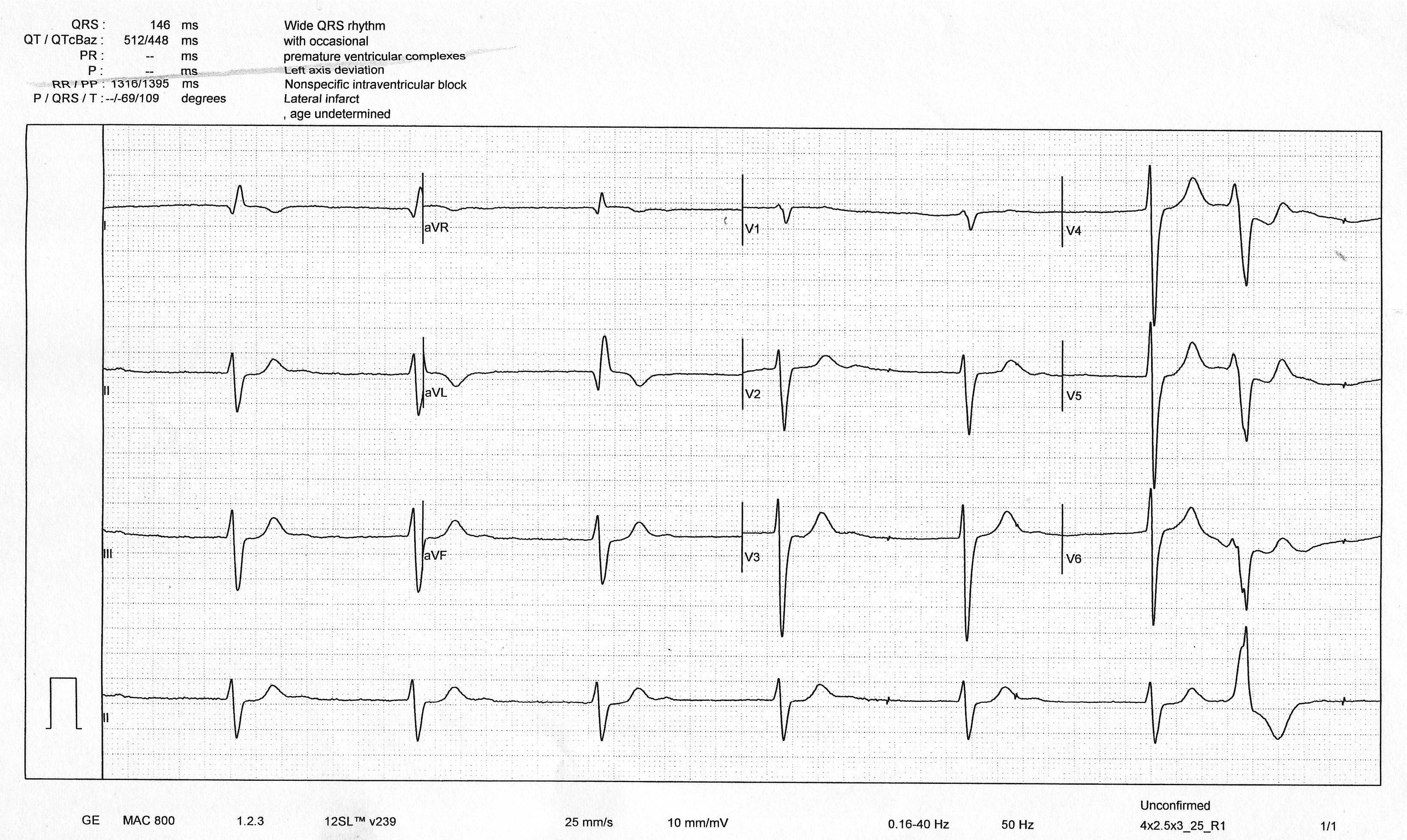
    Normal pacing function of an LV lead.
  • EXPLANATION
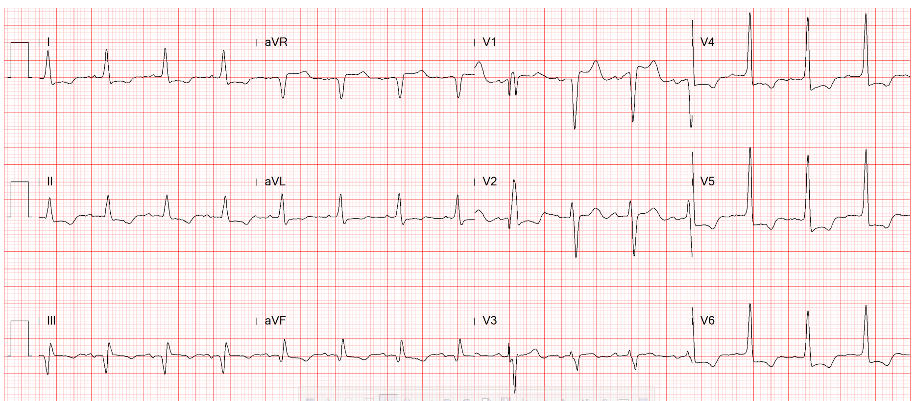
    • Pacing function is normal. Sensing function is unknown.
    • Ventricular pacing vector is left-to-right (positive in V1 and negative in I and aVL) and superior-to-inferior (positive in II, III, and aVF).
    • Pacing has to come from left ventricular side.
    • CXR shows the lead was in outflow tract area. LVOT or CS are possible.
    • X-ray in the LAO view confirms the lead position in coronary sinus.

  • SETTING
    CRT. DDD 60/130.
  • EXPLANATION
    • Atrial rhythm is most likely AF.
    • V pacing is at times very fast.
    • The fastest V pacing is approximately 130 bpm = upper tracing rate.
    • The device is programmed as DDD --> A is tracked. Some atrial signals are not sensed as it is AF.
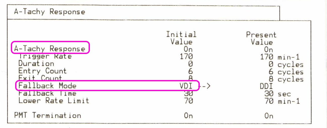
    • If AF is continued to be sensed, DDD mode will be switched to DDI = automode switch algorithm.
    • Conclusion: AF with intermittent rapid V pacing.